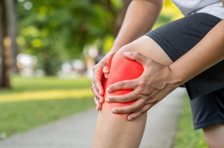
Knee pain can make even simple movements like walking, climbing stairs, or standing up from a chair feel difficult. As one of the body’s major weight-bearing joints, the knee plays a vital role in everyday mobility. At Breeze in Manikonda, Hyderabad, we offer personalised knee pain physiotherapy focused on reducing discomfort, improving stability, and helping you return to active living.

The knee works like a hinge, supported by strong ligaments, muscles, and smooth joint surfaces. When any of these structures are strained, injured, or worn down over time, pain and stiffness can develop. Knee pain may occur suddenly after injury or progress gradually due to joint changes.
Knee pain can develop due to a wide range of reasons, including:
Ligament or cartilage injuries
Meniscus damage
Joint wear and tear over time
Past trauma or fractures
Inflammatory joint conditions
Poor alignment such as bow legs or knock knees
Excess body weight increasing joint stress
Long-standing joint infections or swelling
One of the most frequent causes we see is knee osteoarthritis, a condition where the cushioning between bones gradually wears down. This can lead to pain, stiffness, swelling, and reduced movement, especially during weight-bearing activities.
Pain during walking, standing, or bending
Stiffness after rest or in the morning
Swelling or a heavy feeling in the knee
Grinding or crackling sensation during movement
Reduced ability to fully bend or straighten the knee
A sense of instability or joint locking
Ignoring these signs can affect posture, balance, and overall independence.
At Breeze, Manikonda, Hyderabad, our knee physiotherapy programs aim to:
Reduce pain and swelling
Improve joint flexibility and strength
Support better alignment and movement patterns
Slow down joint degeneration in osteoarthritis
Enhance confidence in daily activities
Our therapists design customised plans using targeted exercises, joint mobility techniques, muscle strengthening, posture correction, and movement education to protect your knees long-term.
Breeze offers compassionate care, experienced physiotherapists, and tailored treatment plans for effective knee pain and osteoarthritis physiotherapy in Manikonda, Hyderabad. Whether your pain is recent or long-standing, we focus on safe recovery and lasting results. Book your appointment today and take the first step toward pain-free movement and stronger knees.